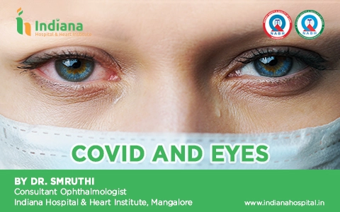
COVID AND EYES
September 6, 2021
By Dr. Smruthi
Consultant Ophthalmologist
Indiana Hospital & Heart Institute, Mangalore

The corona virus disease (COVID-19) caused by the highly-transmissible, acute respiratory syndrome, coronavirus 2 (SARS-CoV-2), has become a global pandemic since December 2019. Initially, there were several reports of irritation in the eye, and eye redness among COVID-19 patients, suggesting that conjunctivitis is an ocular manifestation of SARS-CoV-2 infection.
Indeed, one of the first providers to voice concerns over the spread of coronavirus among Chinese patients was Dr. Li Wenliang MD, an ophthalmologist. He later died of COVID-19, and is believed to have contracted the virus from an asymptomatic glaucoma patient in his clinic.
The prevalence of ocular manifestations among patients with COVID-19 ranges from 2% to 32%.
Ocular manifestations:
A. Eyelid, Ocular Surface and Anterior Segment Manifestations of COVID-19:
1. Follicular conjunctivitis is the most common ophthalmic manifestation documented among COVID-19 patients. Other forms like viral keratoconjunctivitis and pseudomembranous conjunctivitis have also been reported. Patients may have eye redness, ocular irritation, eye soreness, foreign body sensation, tearing, mucoid discharge, eyelid swelling, congestion and chemosis.
Sclera/Episclera: presented as episcleritis.
Anterior Chamber: Acute anterior uveitis has also been reported.
B: Posterior Segment Manifestations of COVID-19:
1. Retina: Etiopathogenesis: Patients of COVID-19 are in a procoagulant state as is evident from an elevated D-dimer, prothrombin time (PT), activated partial thromboplastin time (aPTT), fibrinogen, and cytokines even in the absence of common systemic conditions like hypertension, diabetes or dyslipidemia. Additionally, intermittent hypoxia in patients with pneumonia can induce the endothelial cells to release tissue factor, and trigger a cascade of extrinsic coagulation.a. Central retinal vein occlusion (CRVO), Central retinal artery occlusion (CRAO), Acute macular neuroretinopathy (AMN), paracentral acute middle maculopathy (PAMM).
b. Retinitis, Vitritis and outer retinal abnormalities like Acute Retinal necrosis.
C. Neuro-ophthalmic Manifestations of COVID-19:
1. Papillophlebitis: There may be painless, unilateral, slight diminution of vision. Ophthalmic findings include dilated, tortuous retinal vessels, disc edema, superficial retinal hemorrhages, cotton wool spot with or without macular edema.
2. Optic neuritis: Neurotropism of the virus has been proposed as one of the mechanisms for the neurological and neuro-ophthalmic manifestations. Patients may suffer from painful vision loss, relative afferent pupillary defect (RAPD) in the more severely affected eye with visual field defects and optic nerve enhancement on magnetic resonance imaging (MRI). Treatment is the same as in a typical case of optic neuritis with intravenous methylprednisolone (IVMP) followed by oral prednisolone, leading to visual recovery and resolution of disc edema.
3. Adie’s tonic pupil
A patient with history of retro-ocular pain and difficulty in reading may have a tonic pupil two days after the onset of COVID-19 symptoms.
4. Miller Fisher Syndrome (MFS) and cranial nerve palsy: Onset of acute ataxia, loss of tendon reflexes and ophthalmoplegia, and isolated cases of cranial nerve palsies(3rd, 4th, 6th cranial nerve) have been reported in several patients recently diagnosed with COVID-19. These patients may have a history of acute onset of diplopia.
5. Cerebrovascular accident (CVA) with vision loss: Pre-existing endothelial dysfunction may make patients more susceptible, and they may have onset of acute bilateral, painless loss of vision.
D. Orbital Manifestations of COVID-19:
1. Dacryoadenitis
2. Retro-orbital pain
3. Mucormycosis
Mucormycosis is a life-threatening, opportunistic infection, and patients with moderate to severe COVID-19 are more susceptible to it because of the compromised immune system with decreased CD4+ and CD8+ lymphocytes, and associated comorbidities such as diabetes mellitus, which potentiates both the conditions, and decompensated pulmonary functions and the use of immunosuppressive therapy (corticosteroids).
Rhino-orbital cerebral (ROC) mucormycosis can occur concurrently with COVID-19 infection in patients undergoing treatment, or two to six weeks later. Mortality rate is as high as 50% even with treatment. Early diagnosis with histopathological and microbiological evidence, appropriate management with antifungals and aggressive surgical debridement (functional endoscopic sinus surgery and orbital exenteration) can improve the chances of survival. Simple tests like vision, pupil, ocular motility and sinus tenderness can be part of a routine physical evaluation of a COVID-19 patient hospitalized with moderate to severe infection or diabetics with COVID-19, or those receiving systemic corticosteroids.
A nasal swab for KOH mount and culture is a bedside procedure. Orbital exenteration for life-threatening infection is triaged as an urgent condition requiring surgery within 4-72 hours. Intravenous liposomal amphotericin B is started based on clinical suspicion or results of deep nasal swab.
MRI with contrast is very useful to determine the extent of the disease and intracranial extension. Development of unilateral facial or orbital pain, headache, periocular swelling or double vision, or diminution of vision should prompt even patients who have recovered from COVID-19 to seek immediate medical attention. Since majority of the patients developsymptoms of mucormycosis after recovering from COVID-19, follow-up of high-risk COVID-19 patients for sequelae is imperative.
